Cardiac
Our Specialists
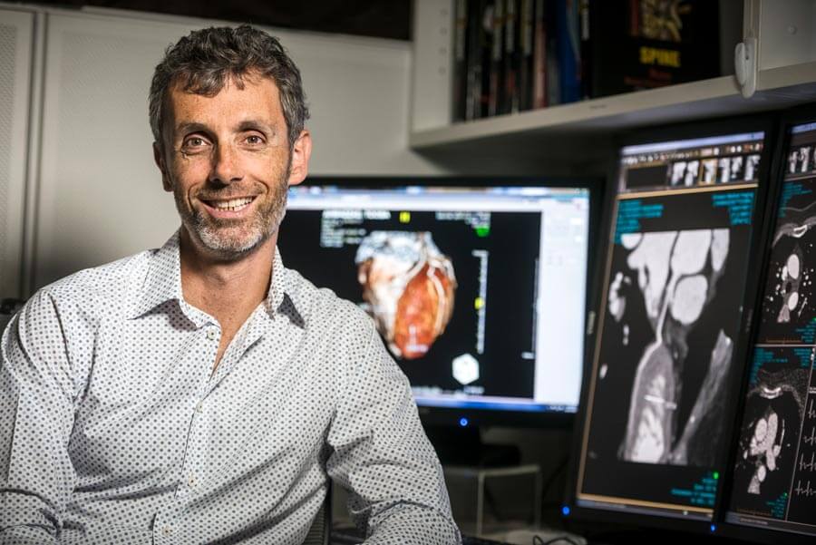
Dr Brendan Adler
Radiologist
Dr Lawrence “Lorry” Dembo
Cardiologist
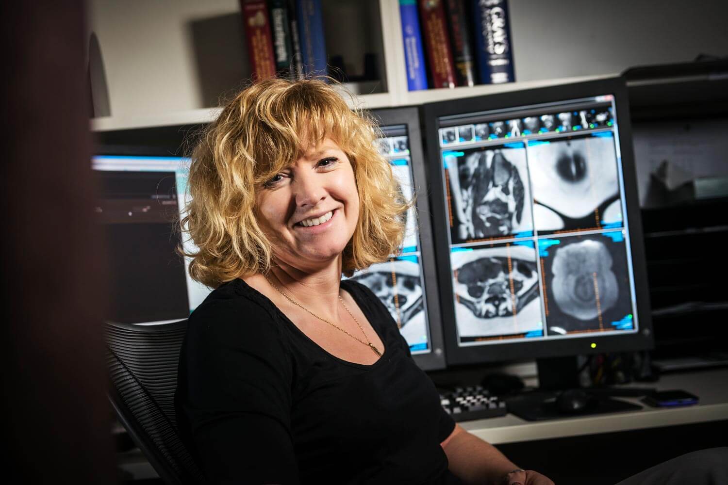
Dr Tonya Halliday
Radiologist
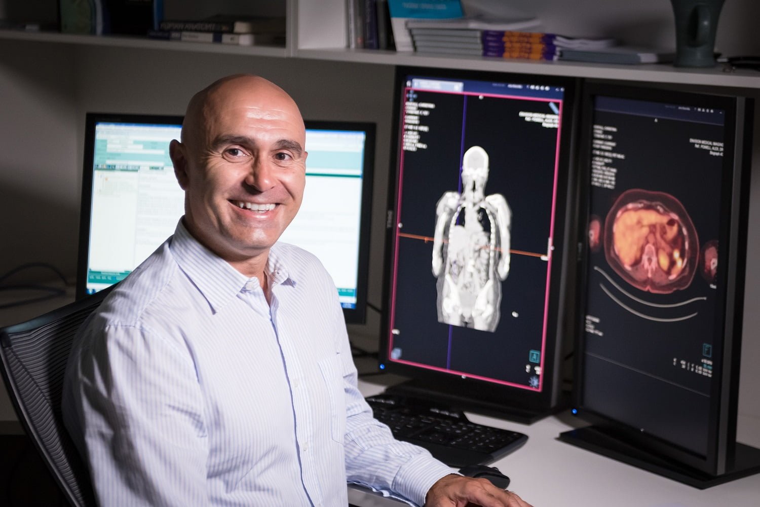
Dr Jerry Moschilla
Radiologist and Nuclear Medicine Specialist
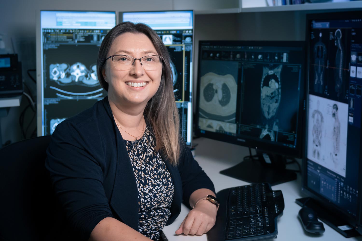
Dr Tracey Muir
Radiologist and Nuclear Medicine Specialist
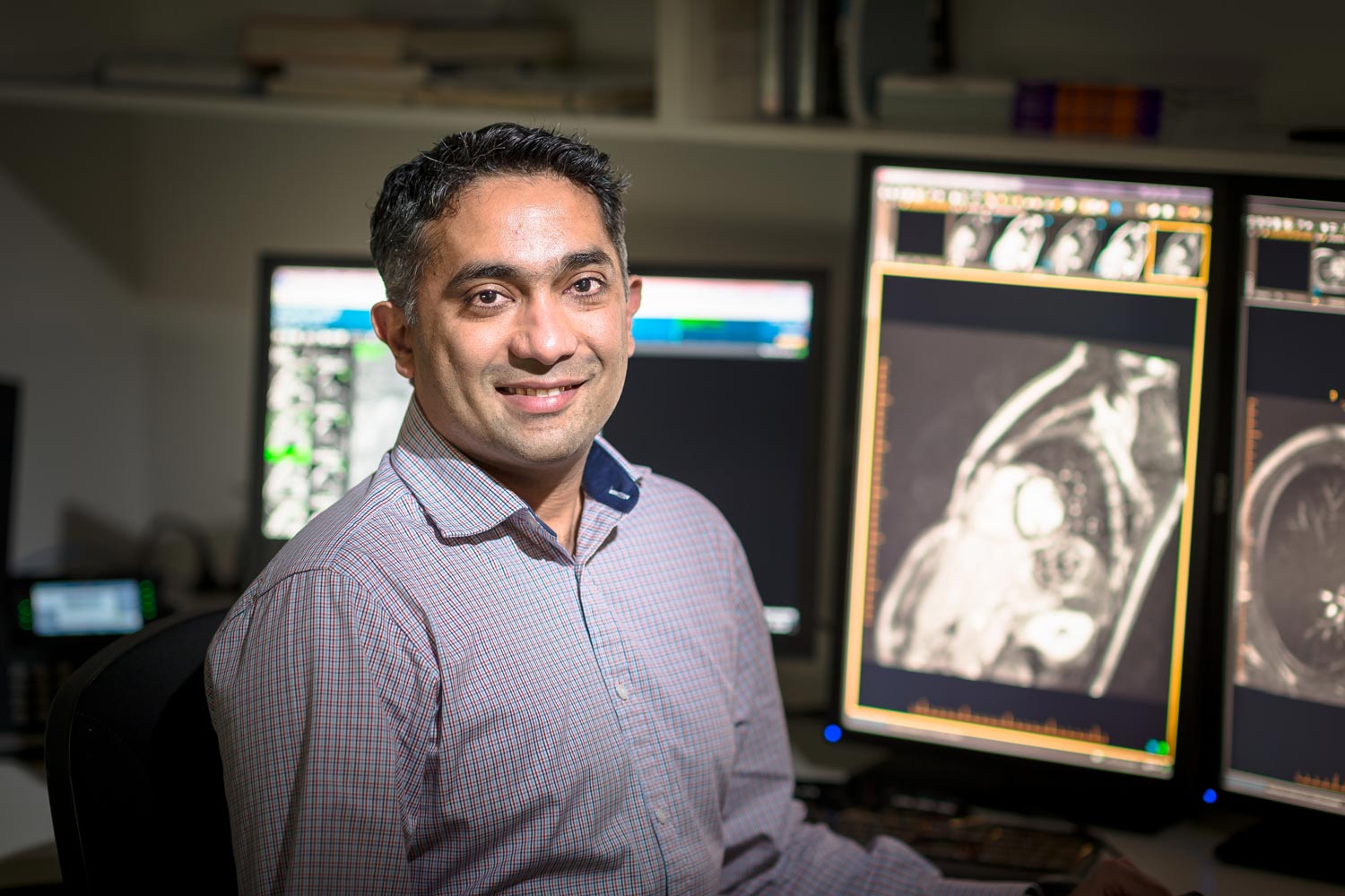
Dr Vikram Raju
Radiologist
Dr Carly Simpkins
Radiologist
Dr Jeanette Soon
Radiologist
Cardiac
Our Services
In comparison to other practices in Western Australia, Envision has, by far, the greatest experience in Cardiac imaging including Cardiac CT, Cardiac MRI and Nuclear Cardiology. Our technology is updated regularly to maintain our world-leading, cutting-edge advantage. We are the preferred provider for the majority of WA cardiologists for a reason – high quality imaging, backed up by experienced, individualized and precise clinical reporting.
Cardiac Imaging at Envision Medical Imaging is a collaboration between Cardiologists, Nuclear Physicians and Radiologists specifically trained in cardiac imaging. Each type of cardiac imaging has its own particular use and is often complimentary to other types of imaging. This multi-modality collaborative approach to cardiac imaging allows comparison and contrast between modalities ensuring the best patient outcomes. Some of our cardiac imaging specialties include:
Nuclear Cardiology
A Myocardial Perfusion Scan images the blood supply (perfusion) to the heart muscle using a gamma camera. The blood supply is made visible to the camera by the introduction of a small amount of radioactive tracer (radiopharmaceutical or MIBI) injected into an arm vein. Occasionally an alternative tracer called Thallium is used. Depending on the exact heart condition in question, the procedure may be performed at rest, with the heart under stress or, most commonly, both. Myocardial scans give information useful in diagnosing and managing conditions such as coronary artery disease, dead tissue resulting from a lack of blood supply (infarcts) and diseases of the heart muscle (cardiomyopathy). A Stress Myocardial Perfusion Scan examines the blood supply (perfusion) to the heart muscle using a gamma camera. The blood supply is made visible to the camera by the introduction of a small amount of radioactive tracer, (radiopharmaceutical or MIBI) injected into an arm vein. The injection is given during stress exercise on a treadmill, then again while resting later on.
After each injection, a gamma camera placed over the chest takes images (maps) the pattern of tracer accumulated in the heart’s left ventricle, giving information useful in diagnosing and managing conditions such as coronary artery disease, infarcts and diseases of the heart muscle – cardiomyopathy.
More Information
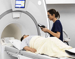
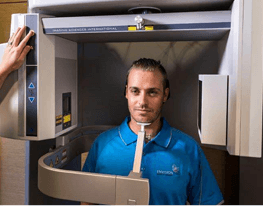
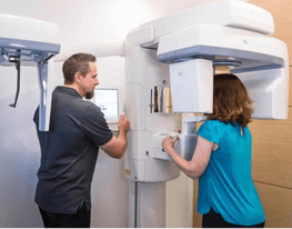
Cardiac CT
CT Coronary Angiography is a specialized form of CT scanning used to take images of the coronary arteries of the heart (angiograms). These arteries supply blood to the heart muscle and disease of these vessels (atherosclerosis) is responsible for most heart attacks.
More Information
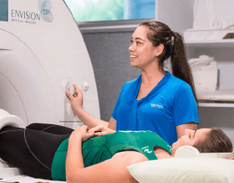
Cardiac MRI
MRI (Magnetic Resonance Imaging) is a way of creating pictures of your body that does not use X-rays or radiation. MRI uses a very powerful magnetic field in combination with a rapidly switching Radio?Frequency (RF) to obtain the images.
MRI is best at demonstrating the soft tissue structures of the body such as muscles, ligaments,the brain and spinal cord. It can also provide unique diagnostic information for Cardiac, Abdominal and Pelvic imaging making MRI advantageous over other imaging modalities for many clinical questions.
More Information
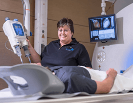
Cardiac
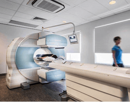
Our Equipment
Our practice is equipped with state-of-the-art digital equipment including 1.5T MRI, 3T MRI, The revolutionary Force multislice CT with cardiac capability, ultrasound, and the latest in filmless digital reporting and image archiving and distribution to ensure the highest level of diagnostic service for cardiac patients.